
Why Is My Dog Limping? Causes, Warning Signs & When to See a Vet
A dog that suddenly starts limping may have a paw injury, sprain, arthritis, torn cranial cruciate ligament, fracture, or another painful condition. In Arizona, veterinarians must also consider Valley Fever when bone pain or unexplained lameness develops.
Desert Forest Animal Hospital evaluates limping and mobility problems in dogs using physical examinations, orthopedic testing, digital X-rays, and laboratory diagnostics when indicated. Our goal is to identify the cause, relieve discomfort, and help owners understand the next step.
Call before arriving. Same-day appointments depend on medical urgency and availability. Major trauma and life-threatening emergencies require immediate emergency veterinary care.
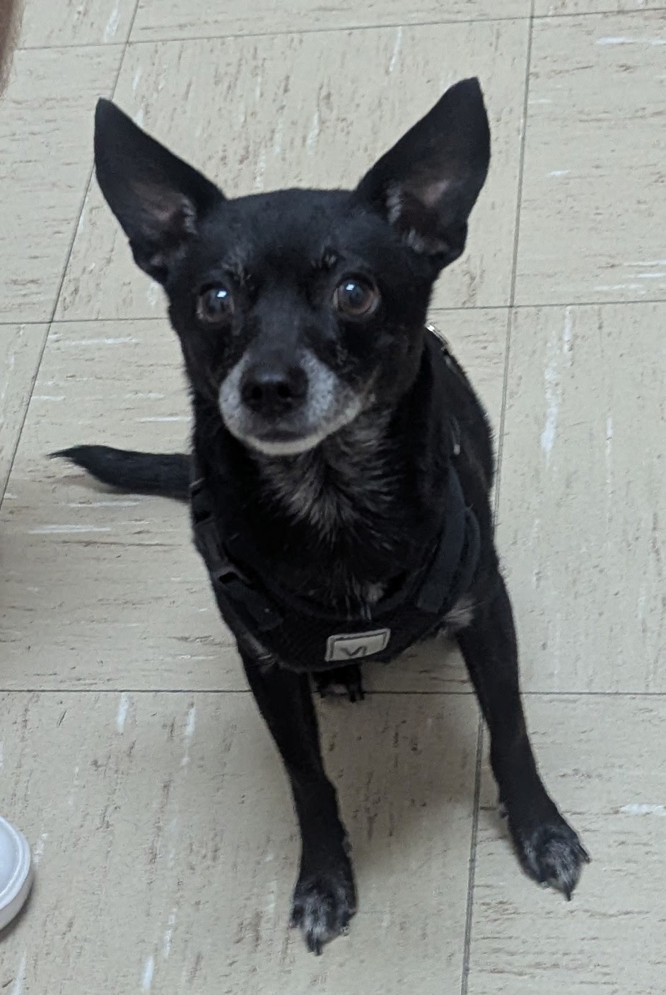
Quick Answer: Why Is My Dog Limping?
Dogs limp when they cannot or will not use a limb normally. Common causes include paw injuries, soft-tissue strains, ligament tears, joint disease, fractures, and infections.
Sudden refusal to bear weight, significant pain, swelling, or worsening symptoms require prompt veterinary attention.
A dog can have a painful injury even without crying, and some serious conditions cause intermittent rather than continuous limping.
When Is Limping an Emergency for a Dog?
Emergency: Seek Care Immediately
- Major trauma or being struck by a vehicle
- Obviously broken or deformed leg
- Inability to stand or walk
- Uncontrolled bleeding or open fracture
- Paralysis or rapidly worsening weakness
- Collapse, severe pain, or breathing difficulty
Urgent: Call a Veterinarian Today
- Sudden refusal to put weight on one leg
- Persistent or worsening new limp
- Pain after running or jumping
- Joint swelling or a painful paw
- Limping with fever or poor appetite
- Suspected cactus spine or puncture wound
Schedule an Examination
- Recurring mild limping
- Stiffness after rest
- Reluctance to jump or climb stairs
- Gradual decline in mobility
- Limping after exercise
- Chronic joint discomfort
A mild limp that resolves quickly may be less urgent than a dog that cannot bear weight. Nevertheless, recurrent or persistent lameness should not be ignored.
Common Causes of Limping in Dogs
1. Paw Injuries, Torn Nails & Foreign Objects
A dog may begin limping after stepping on a cactus spine, thorn, sharp rock, or other object. Torn toenails, pad cuts, burns, and foreign material between the toes can also cause pain.
Check the paw gently if your dog allows it. Do not dig into a wound or attempt to remove deeply embedded objects.
2. Muscle Strains and Sprains
Running, jumping, slipping, and rough play may injure muscles, tendons, or ligaments. A limp that begins after activity is not automatically a simple sprain: fractures and ligament tears may initially look similar.
3. Cranial Cruciate Ligament Tears
The cranial cruciate ligament (CCL) helps stabilize the dog’s stifle, or knee joint. Degeneration, partial tearing, or complete rupture can allow abnormal motion between the femur and tibia, causing inflammation and pain.
Affected dogs may favor a rear leg, touch only the toes to the ground, struggle to rise, avoid stairs, or develop an abnormal sitting posture. Some dogs continue walking despite substantial ligament injury.
Because CCL disease is an important and potentially surgically treatable cause of back-leg limping, it deserves a dedicated orthopedic examination.
4. Osteoarthritis
Arthritis can cause joint stiffness, difficulty rising, reduced endurance, and reluctance to jump. Signs may be especially noticeable after sleeping or lying down.
5. Fractures and Dislocations
Trauma can cause fractures, joint dislocations, soft-tissue damage, and severe pain. Not every fracture causes visible deformity. Diagnostic imaging may be needed to determine the injury.
6. Patellar Luxation
A slipping kneecap may cause a dog to skip, suddenly carry a hind leg, and then resume apparently normal walking. This is common in certain small dog breeds.
7. Hip and Elbow Disease
Hip dysplasia, elbow disease, developmental abnormalities, and secondary arthritis may cause chronic or intermittent limping.
8. Growth-Related Problems
Puppies may develop lameness from growth-plate injuries, developmental orthopedic disorders, inflammatory bone disease, or trauma. Persistent limping in a young dog requires evaluation.
9. Valley Fever and Other Infections
In Arizona, the fungal disease Valley Fever can occasionally spread to the bones and cause pain, swelling, and lameness. Affected dogs may also have coughing, fever, weight loss, poor appetite, or lethargy.
10. Neurologic Disease or Bone Tumors
Spinal and nerve disorders can alter a dog’s gait, while bone tumors and other serious conditions can cause progressive pain or lameness. Foot dragging, knuckling, and loss of coordination require veterinary assessment.
How Veterinarians Diagnose a Torn Cranial Cruciate Ligament (CCL)
CCL disease is one of the most important causes of hind-leg lameness. Diagnosis is based primarily on orthopedic examination and assessment of knee stability, with radiographs providing important supporting information.
Orthopedic Examination: Cranial Drawer and Tibial Thrust
During a stifle examination, the veterinarian evaluates joint pain, swelling, range of motion, muscle condition, and abnormal movement between the femur and tibia.
- Positive cranial drawer sign: The tibia moves abnormally forward relative to the femur when the examiner applies controlled pressure. This indicates instability consistent with CCL rupture.
- Positive tibial thrust (tibial compression test): Flexion of the hock produces abnormal cranial movement of the tibia in a knee with insufficient cruciate ligament restraint.
- Medial buttress: Chronic CCL disease may cause fibrotic thickening along the inner (medial) aspect of the stifle. This is particularly useful as a palpable examination finding.
- Other findings: Joint effusion, decreased range of motion, pain with manipulation, quadriceps muscle atrophy, and possible meniscal injury.
A complete tear often produces obvious joint instability. Partial tears and chronic ligament disease may be harder to detect, particularly when muscle tension or fibrosis restricts movement. Sedation may occasionally be needed for a reliable examination.
What Does a Torn CCL Look Like on a Dog’s X-Ray?
A standard X-ray does not directly show the cranial cruciate ligament. Instead, the veterinarian looks for signs of joint inflammation, instability, and secondary osteoarthritis.
Radiographic findings that may support a CCL injury include:
- Joint effusion: Increased soft-tissue opacity within the stifle, indicating accumulation of joint fluid or inflammatory tissue.
- Compression or effacement of the infrapatellar fat pad: Fluid distending the joint reduces the normal triangular fat-pad shadow in front of the knee joint.
- Distension of the caudal joint capsule: Joint fluid can displace the fat plane behind the knee, another indirect sign of effusion.
- Periarticular osteophytes: New bone growth may develop around the patella, femoral trochlear ridges, tibial plateau margins, and fabellae as osteoarthritis progresses.
- Medial periarticular soft-tissue thickening: Chronic fibrosis associated with a medial buttress may contribute to enlargement of the medial joint silhouette. The buttress itself is primarily assessed by palpation.
- Cranial tibial subluxation: In some cases, the tibia appears displaced forward relative to the femur. This may be more apparent on properly positioned stress radiographs.
- Subchondral sclerosis and enthesophytes: Chronic joint disease may produce increased bone density beneath cartilage and bony proliferation at ligament or tendon attachment sites.
What Happens if My Dog Has a CCL Tear?
Treatment depends on the dog’s size, activity level, medical condition, degree of instability, and severity of joint disease.
Options may include carefully managed activity restriction, pain control, rehabilitation, and surgical stabilization. TPLO is a common surgical option for selected dogs.
Desert Forest Animal Hospital evaluates suspected CCL injuries and discusses appropriate treatment or orthopedic surgical referral.
Learn more about orthopedic evaluations and surgical care at DFAH.
Can Valley Fever Cause a Dog to Limp?
Yes. Valley Fever, also called coccidioidomycosis, is caused by fungal spores found in the desert environment. Dogs become infected after inhaling spores.
The infection commonly begins in the respiratory system. Some dogs develop disseminated disease, meaning the infection spreads beyond the lungs, potentially involving bones, joints, skin, eyes, or other organs.
Bone involvement can cause persistent lameness, localized pain, swelling, or difficulty bearing weight. A dog may have one painful limb or multiple affected areas.
Symptoms That Increase Suspicion for Valley Fever
Unlike an uncomplicated ligament injury, Valley Fever may be accompanied by systemic signs of illness. These can include:
- Persistent or intermittent coughing
- Fever
- Lethargy or reduced activity
- Decreased appetite
- Unexplained weight loss
- Swelling over a painful bone
- Enlarged peripheral lymph nodes
- Joint pain or multiple painful areas
- Occasionally skin, eye, or neurologic abnormalities
Not every dog with Valley Fever has a cough. Some dogs with bone involvement present primarily with lameness or other nonrespiratory signs.
What Does Valley Fever Look Like on a Dog’s Bone X-Ray?
When Valley Fever infects bone, it can produce fungal osteomyelitis, an inflammatory and destructive bone infection.
Possible radiographic findings include:
- Osteolysis (bone destruction): Areas of reduced bone opacity where normal bone has been destroyed by disease.
- Moth-eaten or permeative bone changes: Multiple irregular regions of bone loss may create an aggressive appearance.
- Periosteal new bone formation: Irregular or sometimes substantial new bone may form along the surface of affected bones.
- Mixed lytic and proliferative lesions: Some areas show destructive bone loss alongside reactive bone production. This is a recognized pattern in fungal osteomyelitis.
- Cortical thinning or destruction: Infection may damage the dense outer layer of the bone.
- Adjacent soft-tissue swelling: Inflammation around the affected bone can produce visible surrounding enlargement.
- Single or multiple bone lesions: Disease may involve one bone or several bones, sometimes in different parts of the body.
- Occasional pathologic fracture: Severe bone destruction can weaken the bone enough to increase fracture risk.
Lesions can affect long bones, the pelvis, ribs, vertebrae, and other skeletal structures. The appearance depends on the location and extent of infection.
What Might Chest X-Rays Show in Dogs With Valley Fever?
Because Valley Fever often begins in the lungs, veterinarians may also recommend chest X-rays. Depending on the case, radiographs may reveal:
- Interstitial or nodular pulmonary opacities
- Areas of lung inflammation or consolidation
- Enlarged tracheobronchial or hilar lymph nodes
- Other pulmonary changes consistent with fungal disease
These abnormalities are not specific to Valley Fever, and their absence does not exclude disseminated infection.
Blood Test Findings: Globulins, Monocytes & Inflammation
Blood testing provides important clues when a veterinarian suspects an infectious cause of lameness. Valley Fever can produce abnormalities associated with chronic inflammation, including:
- Hyperglobulinemia: Elevated blood globulin levels may reflect ongoing immune stimulation.
- Monocytosis: Increased monocytes, a type of white blood cell, may accompany chronic inflammation or infection.
- Neutrophilia: Elevated neutrophils may be present with inflammatory disease.
- Hypoalbuminemia: Some affected dogs have decreased serum albumin.
- Other possible findings: Mild nonregenerative anemia or increased alkaline phosphatase, particularly in selected dogs with bone involvement.
These abnormalities are supportive rather than diagnostic. A dog can have Valley Fever without every one of these changes. Elevated globulins or monocytes also occur with other illnesses.
Valley Fever Titers: How the Diagnosis Is Supported
A veterinarian may submit blood for Coccidioides antibody testing, commonly called a Valley Fever test or cocci titer.
The test evaluates whether the dog’s immune system has produced antibodies against the fungus. A positive result, especially when accompanied by compatible clinical signs, radiographic findings, and inflammatory bloodwork, can strongly support the diagnosis.
The laboratory may quantify antibodies as a titer, such as 1:4, 1:16, or 1:32. Higher titers may raise concern about the extent of disease, but the titer does not perfectly predict severity or prove that a bone lesion is caused by Valley Fever.
Results from a reference laboratory commonly take several days, although turnaround varies.
Because results are not always immediately available, the veterinarian may use the examination, bloodwork, and imaging findings to determine safe initial care while awaiting testing.
An early negative test does not completely exclude Valley Fever. Depending on the case, repeat antibody testing after several weeks or additional diagnostic procedures may be necessary.
When confirmation remains uncertain, cytology or biopsy of an appropriate lesion may be recommended to identify fungal organisms or exclude other diseases.
Read our complete Valley Fever guide for Cave Creek dogs and cats.
CCL Tear vs. Valley Fever: How Veterinarians Tell the Difference
A dog with a torn cruciate ligament and a dog with Valley Fever affecting bone may both present with significant hind-leg limping. However, the clinical examination, radiographic pattern, and laboratory findings often provide different clues.
| Finding | CCL Disease | Valley Fever Involving Bone |
|---|---|---|
| History | Sudden or progressive hind-leg lameness; may worsen after activity | Persistent or progressive lameness, sometimes without an obvious orthopedic injury |
| Orthopedic examination | Cranial drawer, tibial thrust, stifle effusion, possible medial buttress | Focal bone pain, swelling, or lameness without characteristic cruciate instability |
| X-ray findings | Joint effusion, compressed infrapatellar fat pad, osteophytes, possible cranial tibial displacement | Bone lysis, irregular periosteal proliferation, cortical destruction, mixed bone lesions, soft-tissue swelling |
| Other symptoms | Usually primarily musculoskeletal; systemic illness is not typical of isolated CCL disease | May include coughing, fever, lethargy, poor appetite, or weight loss |
| Routine bloodwork | No specific diagnostic CBC/chemistry pattern in uncomplicated disease | May show hyperglobulinemia, monocytosis, neutrophilia, or hypoalbuminemia |
| Additional confirmation | Stifle stability testing, orthopedic evaluation, and additional diagnostics when needed | Coccidioides serology interpreted with clinical findings; sometimes cytology or biopsy |
| Treatment approach | Pain management, controlled activity, rehabilitation, or orthopedic surgery | Veterinarian-directed antifungal treatment and monitoring, often over many months |
Why This Matters for Dogs in Arizona
A dog with a painful back leg should not automatically be diagnosed with a cruciate tear, and a dog with an abnormal bone X-ray should not automatically be diagnosed with Valley Fever.
The combination of orthopedic examination, radiographs, CBC/chemistry findings, exposure history, and targeted laboratory testing helps guide an accurate diagnosis.
How DFAH Evaluates a Dog With Unexplained Limping
A thorough evaluation begins with understanding when the lameness started, whether trauma occurred, how the dog bears weight, and whether additional symptoms are present.
Step 1: Physical and Orthopedic Examination
The veterinarian observes gait, assesses muscle condition, palpates the affected limb, checks joint motion, and evaluates for swelling, instability, or neurologic changes.
Step 2: Digital Radiographs When Indicated
X-rays may help identify fractures, arthritis, joint effusion, bone destruction, or other clinically important abnormalities.
Standard radiographs are particularly useful when a bone or joint problem is suspected, although they do not directly visualize every ligament, tendon, or nerve.
Digital X-Rays in Cave Creek
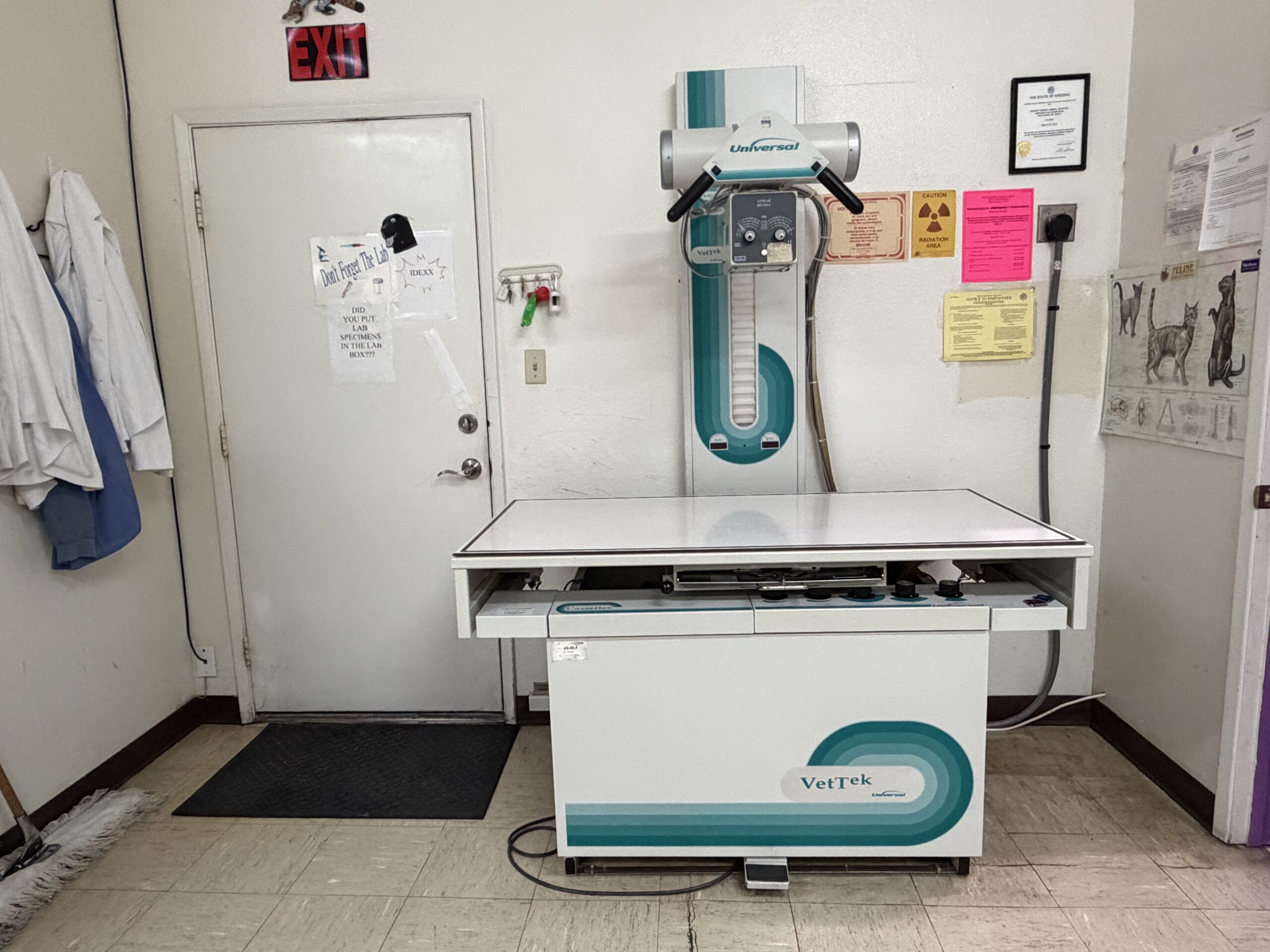
Desert Forest Animal Hospital provides in-house digital radiography for dogs and cats.
Imaging can help distinguish a suspected orthopedic problem from a bone lesion that may require infectious-disease testing or further evaluation.
Learn more about our veterinary X-ray and diagnostic imaging services.
Step 3: Bloodwork and Infectious-Disease Testing
When systemic disease is suspected, a complete blood count, serum chemistry panel, and targeted infectious-disease testing may be recommended.
For a dog with compatible clinical findings in Arizona, Valley Fever serology may be appropriate. Testing is not required for every dog that limps.
Step 4: Treatment or Referral Planning
The veterinarian discusses the likely diagnosis, appropriate pain control, recommended activity, further testing, and whether specialized treatment or referral is necessary.
How Is Limping Treated in Dogs?
Treatment depends on the diagnosis. Options may include rest, prescription pain management, wound care, rehabilitation, joint disease management, orthopedic surgery, or treatment of an underlying infectious condition.
Rest and Activity Restriction
Some injuries require temporarily restricting running, jumping, stairs, and rough play. The duration and intensity of restrictions should be based on the injury.
Veterinary Pain Management
Your veterinarian may prescribe medication appropriate to your dog’s age, medical history, kidney and liver function, and current condition.
Do not administer ibuprofen, naproxen, acetaminophen, or other human pain medicines without explicit veterinary instructions. These medications can cause serious harm to pets.
Arthritis and Long-Term Mobility Care
Dogs with osteoarthritis may benefit from appropriate weight management, controlled exercise, veterinary medications, rehabilitation, and home environment adjustments.
Orthopedic Stabilization
Dogs with clinically important CCL instability or certain fractures may need specialized surgical treatment. DFAH can discuss appropriate surgical options and referrals.
Treating Valley Fever
Dogs with clinically significant Valley Fever generally require veterinarian-directed antifungal treatment and follow-up. The treatment course may last many months, particularly when the bones or other extrapulmonary tissues are involved.
Follow-up may include monitoring clinical signs, bloodwork, serology, and radiographs when indicated.
What Should I Do at Home If My Dog Starts Limping?
- Limit activity. Avoid running, jumping, and rough play.
- Inspect the paw gently. Look for torn nails, visible wounds, or superficial debris. Do not force a painful exam.
- Do not manipulate the painful leg. Avoid repeatedly bending or stretching the joint.
- Record a short video if your dog can walk comfortably and safely. Do not force a dog to walk for a video.
- Avoid human pain medications. Call your veterinarian for medication guidance.
- Contact your veterinary team. Describe the affected leg, onset, weight-bearing ability, and any recent injury or systemic symptoms.
Do not delay care for severe pain, inability to stand, major trauma, or rapidly worsening symptoms.
Why Choose Desert Forest Animal Hospital for a Limping Dog?
Desert Forest Animal Hospital is an independently owned veterinary practice in Cave Creek, Arizona. We provide individualized examinations and diagnostic services for dogs with pain, injuries, and mobility concerns.
Orthopedic Examinations
Evaluation of joint instability, muscle and ligament injuries, fractures, and chronic mobility problems.
In-House Digital X-Rays
Imaging to evaluate bones and joints when indicated, with additional diagnostic recommendations based on clinical findings.
Arizona-Specific Diagnostics
Experience evaluating local conditions including desert paw injuries and Valley Fever.
We serve pet owners from Cave Creek, Carefree, Desert Mountain, Terravita, Dove Valley Ranch, Tatum Ranch, Desert Hills, Anthem, and North Scottsdale.
Related services: same-day urgent care, digital veterinary X-rays, orthopedic surgical evaluations, and Valley Fever diagnosis and treatment.
Frequently Asked Questions
Can a torn CCL be diagnosed without an X-ray?
Often, an orthopedic examination demonstrating cranial drawer or tibial thrust can establish clinically significant stifle instability. Radiographs provide supporting information, help exclude other problems, and may be needed for surgical planning.
What does joint effusion look like on a dog’s knee X-ray?
Joint effusion can increase soft-tissue opacity around the stifle and compress the normal infrapatellar fat-pad shadow. Distension of the caudal joint capsule may also be visible.
What is medial buttress in a dog with a CCL tear?
Medial buttress refers to fibrotic thickening along the inside of the stifle, often associated with chronic CCL instability. It is primarily a finding on palpation, although related soft-tissue enlargement can sometimes be appreciated on radiographs.
Can a partial CCL tear have a negative drawer test?
Yes. Partial tearing, muscle tension, and chronic joint fibrosis can reduce detectable instability. A veterinarian may use additional examination techniques, sedation, radiographs, or further diagnostics when needed.
Can Valley Fever cause limping without coughing?
Yes. Bone involvement can cause lameness or pain even when respiratory symptoms are absent or not obvious. A veterinarian considers the entire clinical picture.
What bone changes can Valley Fever cause on X-rays?
Valley Fever osteomyelitis may cause bone destruction, irregular new bone formation, cortical damage, mixed lytic-proliferative lesions, and soft-tissue swelling. These changes are not specific to the fungus.
Do high globulins and monocytes mean my dog has Valley Fever?
Not necessarily. Hyperglobulinemia and monocytosis can accompany Valley Fever, but both are nonspecific findings. Other infectious, inflammatory, and medical conditions can cause similar changes.
How long does a Valley Fever titer take to return?
Reference-laboratory testing commonly takes several days. The exact turnaround depends on the laboratory, test method, shipping, and processing schedule. Your veterinarian can explain the expected timeframe.
Does a positive Valley Fever titer confirm active disease?
A positive result indicates antibodies to Coccidioides and strongly supports the diagnosis in a dog with compatible symptoms and other findings. However, some exposed dogs may have positive antibodies without active clinical disease. Further evaluation may be necessary.
Does a negative Valley Fever test rule out infection?
No. Tests may be negative early in infection or in some seriously ill dogs. Repeat testing after several weeks or other diagnostic methods may be necessary when suspicion remains high.
Does my dog need an X-ray for every limp?
No. Imaging decisions depend on the history, examination, severity, and suspected underlying problem. Some diagnoses rely primarily on examination, while others benefit from radiographs or additional testing.
Can DFAH see my dog the same day?
Same-day urgent evaluations may be available during regular business hours. Please call (480) 488-2010 before arriving. Major trauma and other critical emergencies may need a 24-hour emergency hospital.
Dog Limping? Call Our Cave Creek Veterinary Team
Whether your dog has a sudden back-leg limp, an injured paw, recurring stiffness, or unexplained bone pain, our team can help determine the appropriate next step.
Desert Forest Animal Hospital
6554 E Cave Creek Road
Cave Creek, AZ 85331
Regular Hours:
Monday-Friday: 9 AM-6 PM
Saturday-Sunday: Closed
Call ahead for urgent concerns. Same-day availability is not guaranteed. Seek immediate emergency care for major trauma, severe injury, inability to stand, or life-threatening illness.
Veterinary Medical References
The diagnostic guidance above draws on veterinary surgical, internal medicine, and infectious-disease resources.
- American College of Veterinary Surgeons: Cranial Cruciate Ligament Disease
- Today’s Veterinary Practice: CCL Diagnosis and Radiographic Findings
- University of Arizona: Valley Fever Diagnosis in Dogs
- Merck Veterinary Manual: Coccidioidomycosis in Animals
- Journal of the American Veterinary Medical Association: Coccidioides Osteomyelitis in Dogs
- U.S. FDA: Pain Medication Safety for Pets
Veterinary patient-education notice: This resource provides general information and does not replace a veterinary examination. Diagnosis and treatment must be individualized.